TMJ Surgery Before and After Case Studies
TMJ Surgery #1: TMJ Osteoarthritis
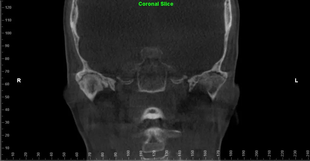
This patient developed severe TMJ osteoarthritis bilaterally. This resulted in pain, limited function, dietary restrictions disrupting her quality of life. She required bilateral TMJ total joint replacement and Lefort I osteotomy to treat the TMJ disease, malocclusion, and facial esthetics.


TMJ Surgery #2:
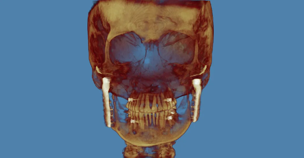
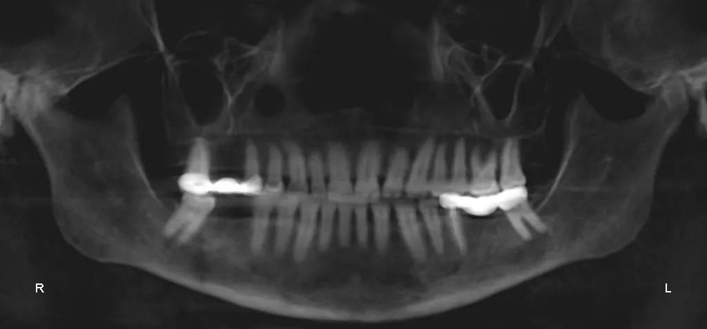
This patient developed debilitating bilateral TMJ osteoarthritis resulting in pain, limited function, and decreased quality of life. She was treated with bilateral TMJ total joint replacement. Note the minimal appearance of her incisions at 6 weeks after surgery.


TMJ Surgery #3
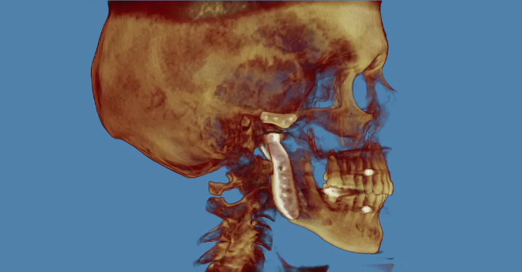
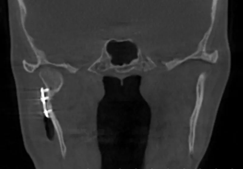
This patient developed heterotopic (excess) bone formation around her right TMJ. This resulted in minimal mouth opening, pain, and limitations to her diet. She was treated via removal of the heterotopic bone, right TMJ total joint replacement, and abdominal fat grafting to avoid further bone formation.


TMJ Surgery #4
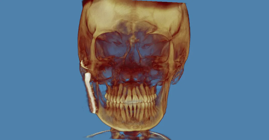
This patient developed heterotopic bone formation following previous TMJ surgery which resulted in severe limitations to mouth opening, severe pain, and significant disruption of her quality of life. She was treated with bilateral TMJ Total Joint Replacement with abdominal fat graft harvest.


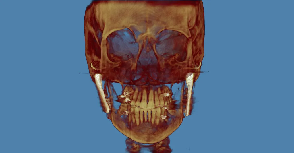

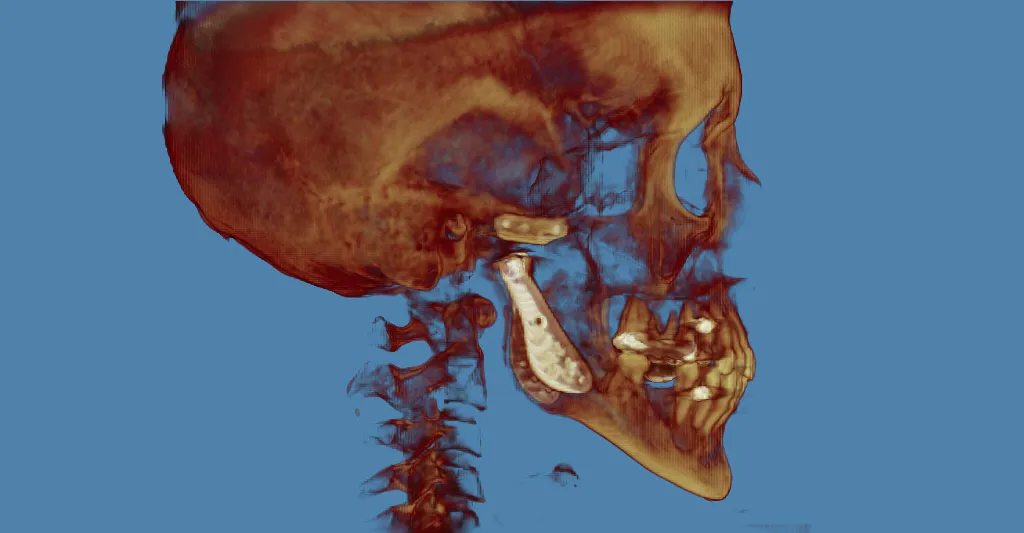

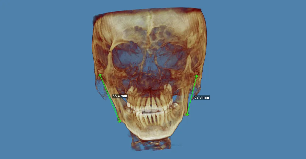
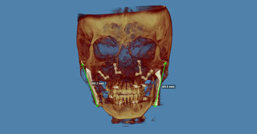
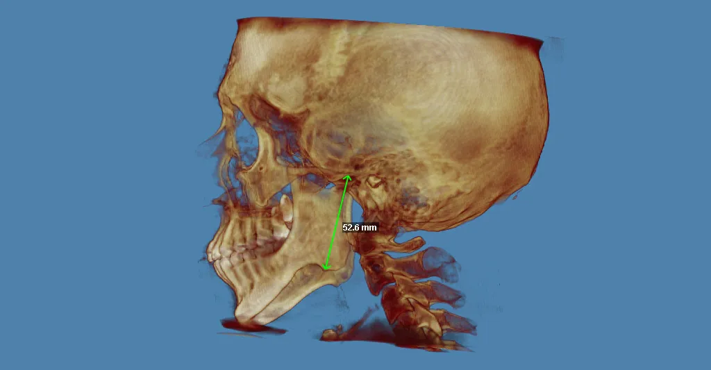
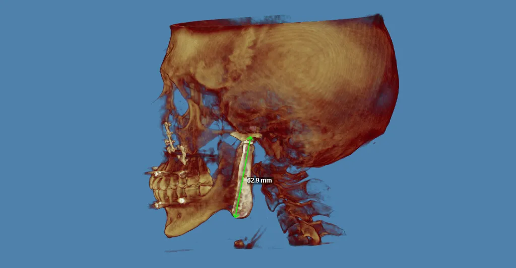
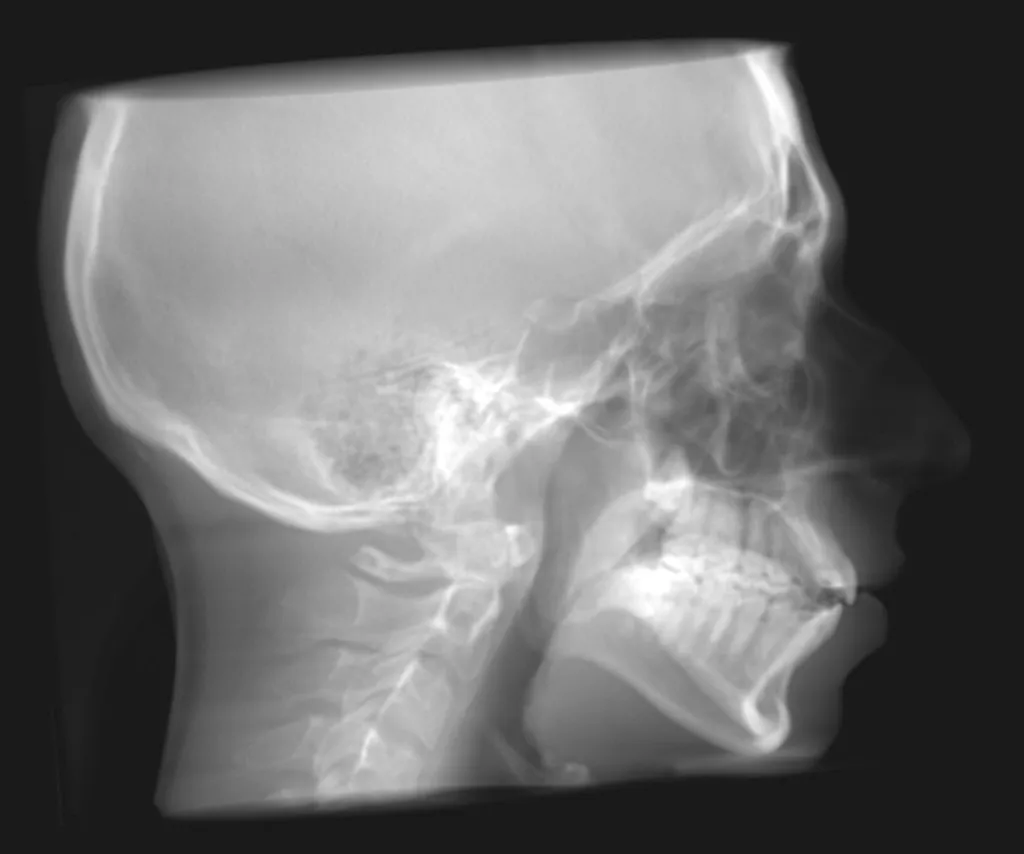
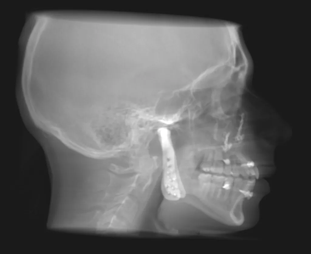
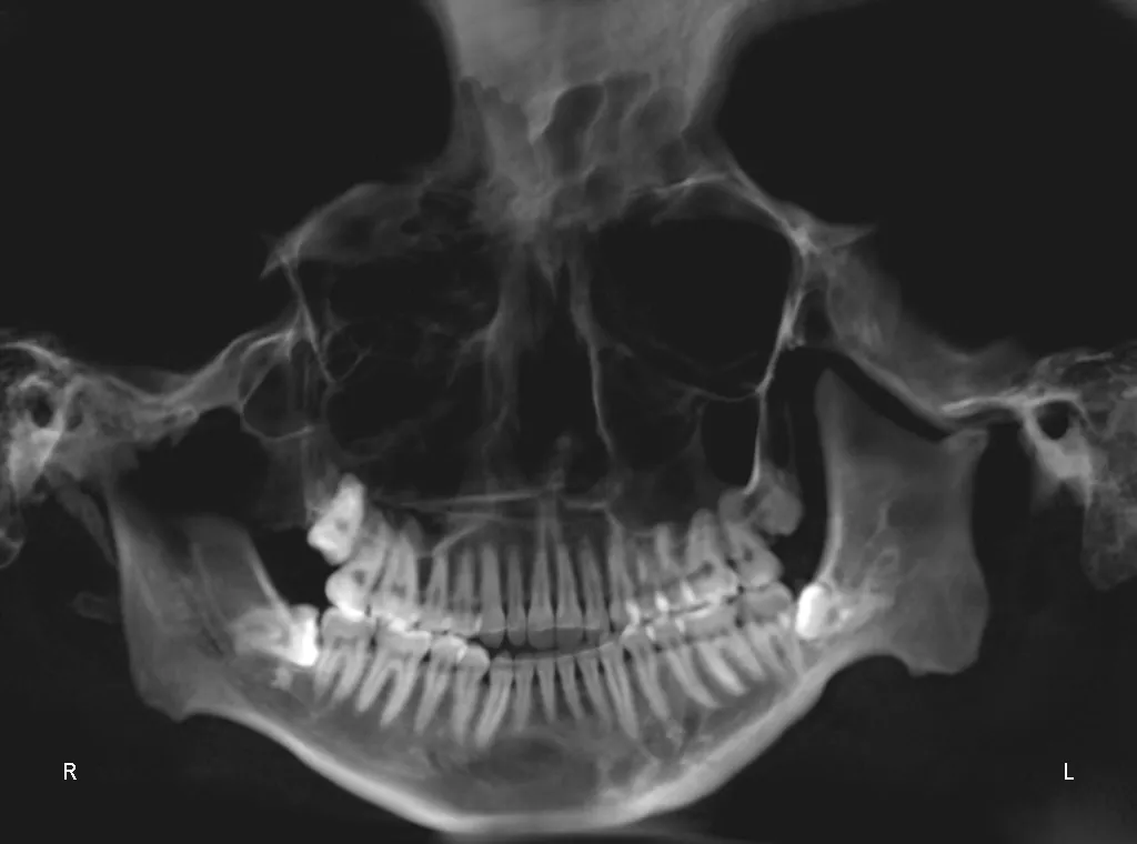
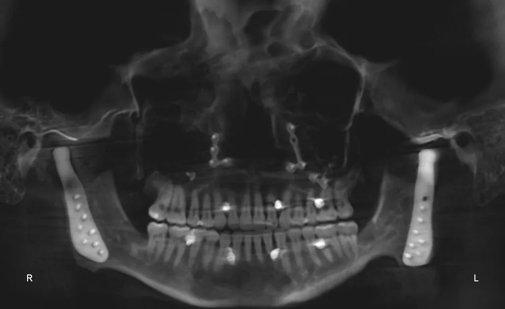
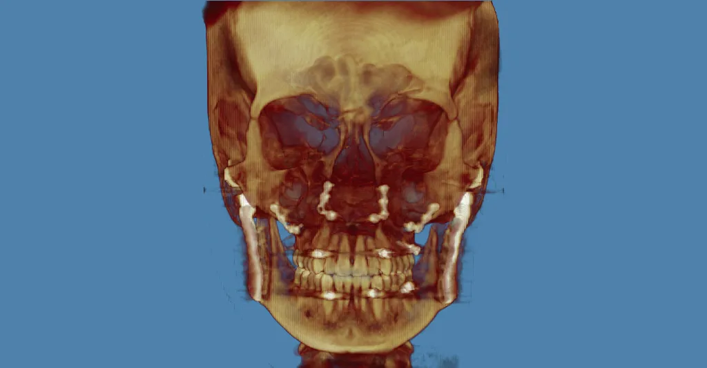
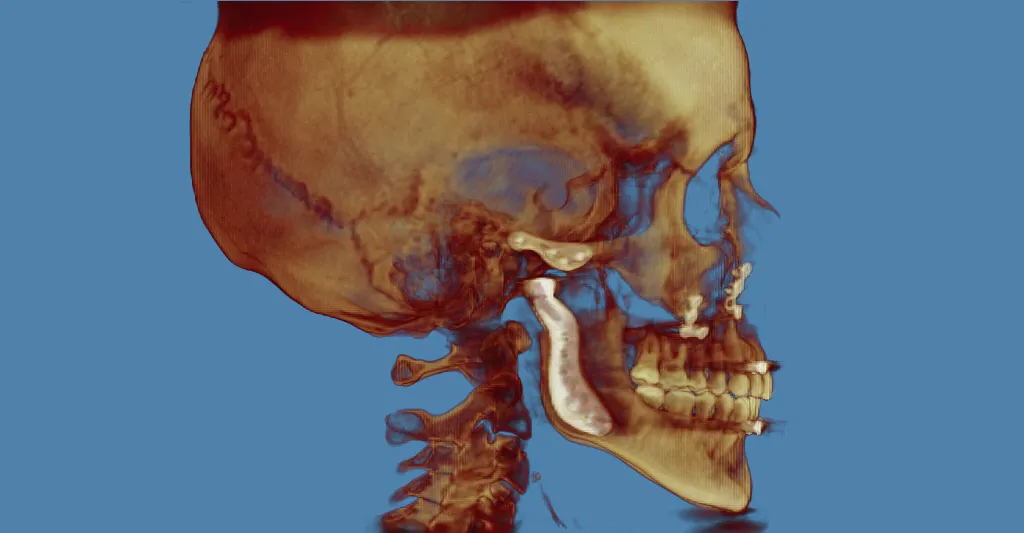
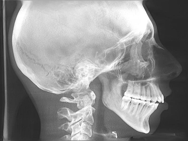
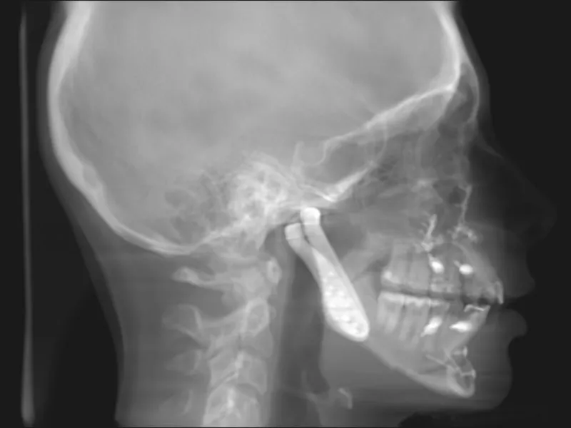
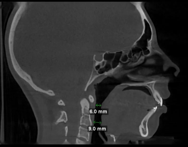
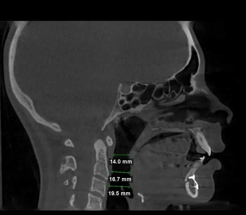
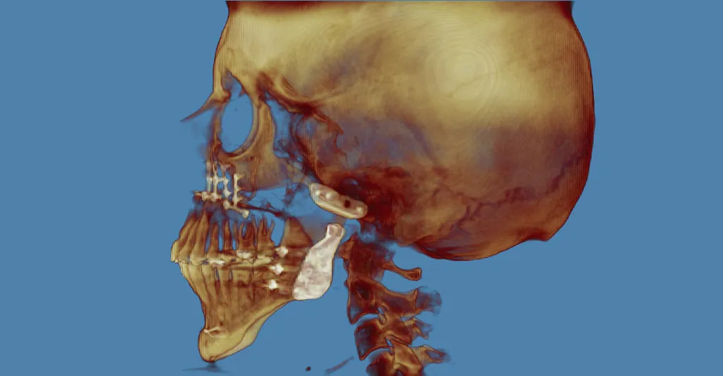
TMJ Surgery #5: TMJ Total Joint Replacement with Orthognathic Surgery
This 22 year old patient had Idiopathic Condylar Resorption (ICR), anterior open bite malocclusion, and a restricted airway. Following Bilateral TMJ total joint replacement and segmental Lefort I osteotomy she has an improved occlusion, a much larger airway (see images), and improved function. She can open to 43mm and bite into foods with her front teeth for the first time in years.


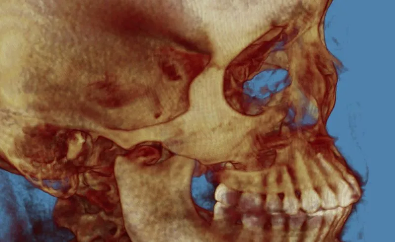
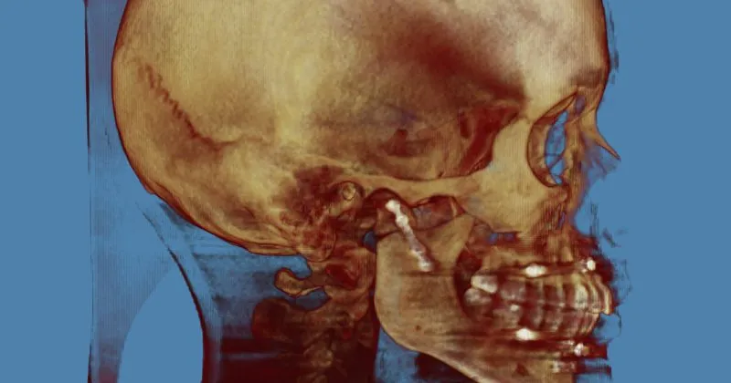
TMJ Surgery #6: Revision Jaw Surgery
This patient had a right subcondylar fracture of the mandible that was treated by another surgeon with Maxillomandibular Fixation (MMF or jaws wired shut). This resulted in malocclusion and decreased ability to move the mandible. After revision surgical treatment she can open, chew normally, and has a return to her pre-injury occlusion (bite).


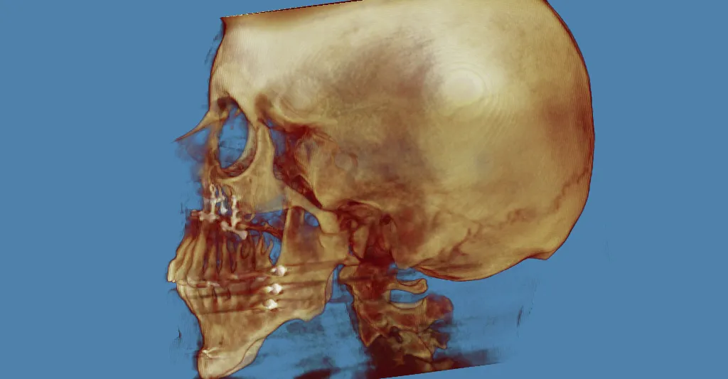
TMJ Surgery #7: Revision Jaw Surgery
This patient had a fall that resulted in a subcondylar fracture of the right mandible. She was treated by another surgeon with Maxillomandibular Fixation (MMF or mouth wired shut). This resulted in malocclusion with limited function. She was treated with revision jaw surgery resulting in return of her normal occlusion and excellent function.


TMJ Surgery #8: Rare Tmj tumor
This patient had a rare TMJ tumor known as synovial chondromatosis which was diagnosed with a biopsy during advanced TMJ surgical arthroscopy. As seen in the video link below, the TMJ was filled with fragments of cartilaginous tissue causing pain, limited mouth movement, and occlusal changes. The tumor was removed in its entirety with an open arthrotomy into the TMJ. The fragments can be seen in the photos below showing extrusion of the tumor in the surgical image, and their removal in the subsequent image.


TMJ Surgery #9:
This patient had an accident as a child causing heterotopic bone formation and ankylosis resulting in near fusion of her TMJ to her skull base. After decades of limited mouth opening and minimal function, we resected the excess bone and performed TMJ total joint replacement. Prior to surgery, the patient’s mouth opening was less than 15mm and had difficulty eating normally. Following surgery, the patient can now open to 37mm and eat normally.


TMJ Surgery #10
This young patient had condylar degeneration (likely Idiopathic Condylar Resorption) resulting in anterior open bite malocclusion, decreased airway space, and decreased mandibular function. We performed bilateral TMJ total joint replacement and lefort I osteotomy to restore mandibular function, correct the malocclusion, improve facial esthetics and improve the airway.